 We developed a system for quantitative imaging of total cytoplasmic glutathione (GSH) in vivo following GST-catalysed conjugation to monochlorobimane (MCB) to give a fluorescent glutathione-bimane (GSB) adduct. This has allowed measurement of concentrations of GSH in defined cell types in intact tissues, to dissect the control of GSH synthesis and to analyse activities of GSH-based xenobiotic detoxification pathways in intact tissues with sub-cellular resolution.

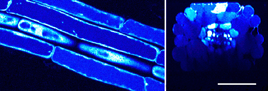
The imaging assay is technically complex (Fricker et al., 2000; Meyer and Fricker, 2000; Meyer et al., 2001), but has now been successfully applied to GSH measurements in several different types of tissues including roots (Sanchez-Fernandez et al., 1997; Fricker et al., 2001), trichomes (Gutiérrez-Alcalá et al 2000), suspension culture cells (Meyer and Fricker, 2002), mesophyll, epidermal and guard cells of wild type and transgenic poplar over-expressing γ-ECS (Hartmann et al 2003), mutants in ER morphology (Au et al., 2012), and most recently pathogenic fungi (Samalova et al., 2014).
