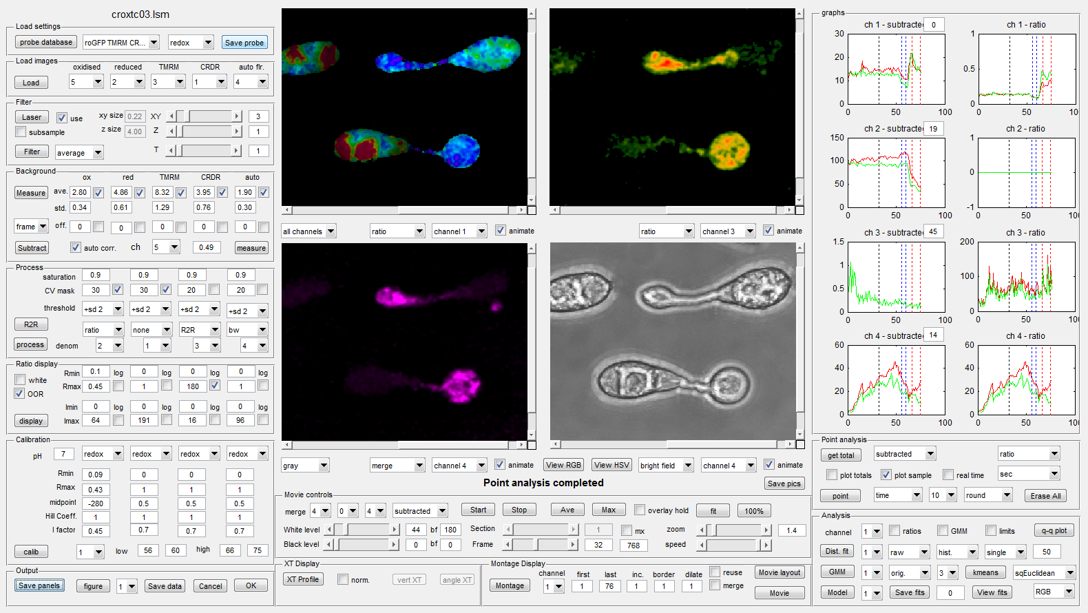
Advance Ratio Analysis
The Advanced Ratio Program extends the basic ratio analysis to handle 4-D (x,y,z,t) images with up-to 5 fluorescence channels, including an auto-fluorescence channel,, and parallel bright field images. This allows correlation of changes in auto-fluorescence-corrected ratio images with up to two other physiological parameters, and (non-confocal) bright-field morphology. In addition to the manual ROI measurements, movies and montages available in the basic ratio analysis module, there is also an x-t kymograph option to represent the response along a manually defined transect as a 2-D image with response on the x-axis and time on the y-axis.
Pixel-population measurements
In more complex images, such as intracellular hyphal networks during fungal infection (13), picking a few ROIs across the specimen does not provide a robust, un-biased estimate of the physiological behaviour throughout the system. However, this can be achieved in the advanced module by considering the intensity or ratio information from a statistical perspective along with estimates of the goodness-of-fit, and either calculating multi-component 1-D Gaussian fits to the log ratio data from all pixels or fitting 2-D Gaussian Mixture Models (GMM) to the corrected intensity data prior to ratioing. Fitting to the original intensity data or log ratio values does not take into account the relative intensity of the different pixels contributing to the distributions. Thus ratios from very dim pixels contribute as equally as ratios from very bright pixels. A better alternative is to calculate a 1-D or 2-D weighted histogram in which each data point is replicated in the data set in proportion to the average intensity at each of the two wavelengths. This ensures that a large number of dim and noisy pixels do not skew the fits. The advanced ratio module includes options to explore these data fitting approaches.
